


With the rapid growth of personalized medicine, there is a growing need for innovative manufacturing technologies. Additive manufacturing (AM) technologies are highly regarded for their unique manufacturing advantages and are particularly valuable in the field of tissue engineering. In particular, melt electrostatic spinning (MEW) technology has attracted much attention for its ability to precisely fabricate 3D microfibrous scaffolds with controllable, bionic micro- and nanostructures. However, MEW technology faces a number of technical challenges when constructing scaffolds with hollow regions or suspended structures, as required for vascularization applications. To overcome these limitations, this study developed microfibrous scaffolds with branched hollow structures by innovatively combining fused deposition molding (FDM) and MEW technologies, which exploits the complementary advantages of both technologies while significantly expanding their potential applications in tissue engineering.
01|Materials And Methods
a.Material Selection
Conductive polylactic acid (cPLA) was chosen as the mold material for FDM printing in the study, with a resistivity of about 0.7 Ω-m, which is close to that of seawater (0.223 Ω-m) and shows good electrical conductivity, compared to copper's resistivity of only 16.8 nΩ-m. Polycaprolactone ( PCL) as the biomaterial for MEW, as it is not only compatible with MEW technology, but also approved by the U.S. Food and Drug Administration (FDA) for use in Class III medical devices (including long-term implants) due to its excellent biocompatibility.
b.Mold Design And Preparation
A mold half with an arterial branching pattern was designed using the computer-aided design (CAD) software SOLIDWORKS, as shown in Fig. 1, where the radius of the mold was gradually reduced from 5 mm at the bottom to 2 mm at the farthest end of the branch, with an additional 2-mm filler layer at the bottom to facilitate the subsequent fusion process of the stent.
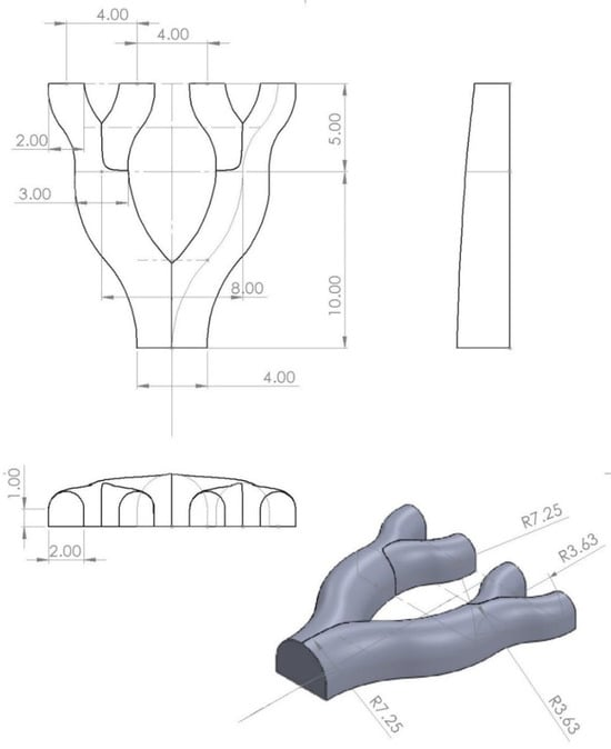
Fig 1. Schematic diagram of the branching mold (unit: mm)
c.Fusion Electrophotographic Printing Process
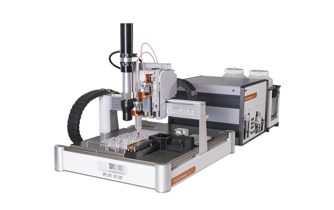
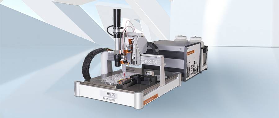
The cPLA molds, pre-printed via FDM technology, are placed on a glass printing table above the high-pressure platform of the Gesim BioScaffolder 3.1 device. It is worth highlighting that the Gesim BioScaffolder 3.1 is a state-of-the-art 3D bioprinting platform equipped with multiple independent print nozzles in the Z-axis direction, which supports the mixing and combining of multiple materials for printing in the same print case, an advantage that can meet the requirements of manufacturing complex bionic structures. During the printing process, the nozzles are maintained at a height of a few centimeters above the mold surface, ensuring precise alignment with the mold. After optimizing the fused electro-writing printing parameters, the PCL material was used to print on the cPLA mold. This method can effectively control the flow rate of the fused filament and avoid material buildup at the edges of the holder. The optimized MEW process parameters are shown in Table 1.

Table 1. Optimized MEW printing parameters
d.Post-processing And Assembly Of Brackets
After the fusion electrowriting printing was completed, the newly made MEW scaffolds were carefully removed from the cPLA mold. Subsequently, the two corresponding MEW scaffold halves were placed on a heated platform with a set temperature of 85°C for hot-melt merging and cooled at room temperature to successfully prepare the hollow branching scaffold structure as shown in Figure 2.
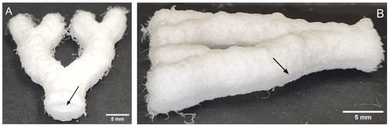
Fig. 2. Complete three-dimensional hollow branching stent structure. (A) Arrows indicate hole openings. (B) Arrows indicate heat-fused longitudinal joints
Through the above steps, hollow microfiber scaffolds with complex branching vascular structures were successfully prepared by combining the advantages of both FDM and MEW additive manufacturing technologies.
a.Mold Manufacturing Results
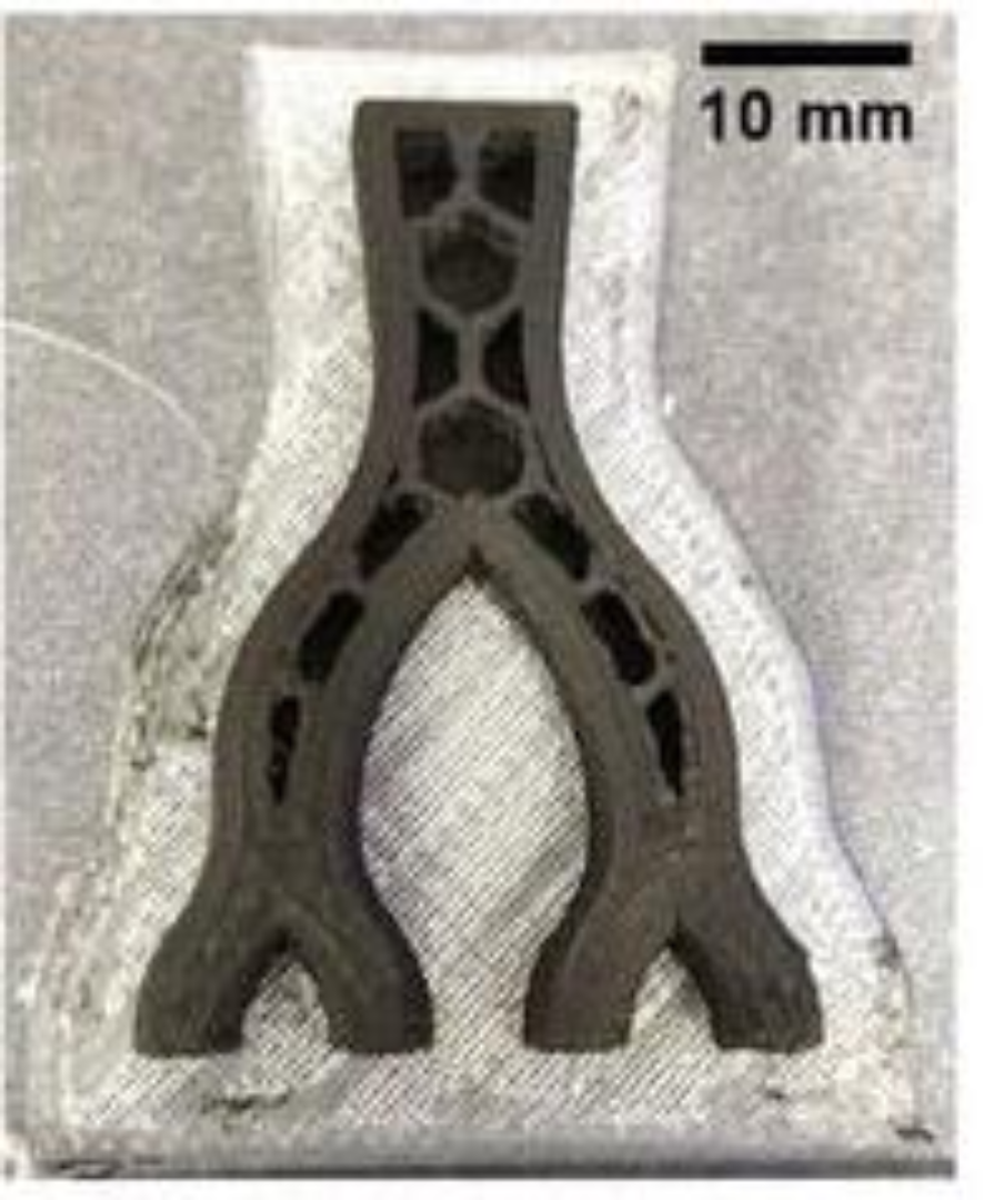
Using the FDM technique, a graphene-added conductive polylactic acid (cPLA) mold was successfully fabricated, as shown in Figure 3. Although cPLA exhibited some brittleness, this did not negatively affect the overall quality of the mold. It is worth mentioning that the cPLA mold exhibited good interfacial compatibility with the final melt electro-written (MEW) scaffolds, and the scaffolds were able to be easily removed from the molds without being damaged.
b.MEW Print Quality Analysis
Compared to glass molds, MEW scaffolds fabricated on cPLA molds have smaller pore sizes and finer fiber diameters, as shown in Fig. 4. cPLA molds have fiber diameters of about 30 μm, compared to 35 μm on glass molds.
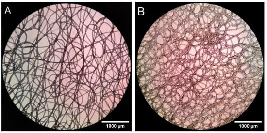
Fig 4. single-layer MEW holder printed on glass (A) and cPLA mold (B)
c.Pore Characterization
The porosity was estimated to be about 63.29% and the average pore diameter was about 30 μm by the body-visualization method (Table 2), which is in general agreement with the result of 77.08% measured by the relative density method.
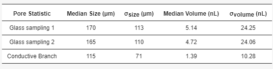
Table 2. Mean pore size and volume estimated by stereology
d.Mechanical Characterization
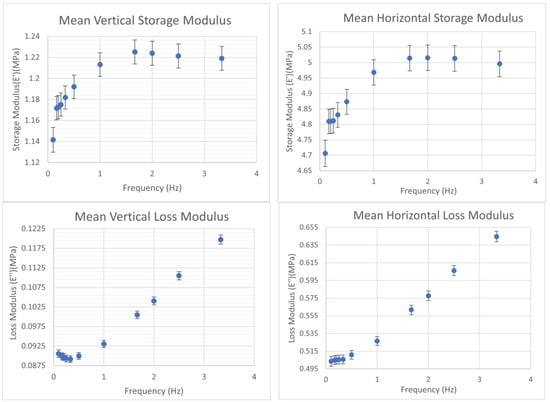
The results of dynamic mechanical analysis (DMA) showed that the prepared MEW scaffolds displayed significant anisotropic mechanical properties at 37.5°C, as shown in Fig. 5. Under the compressive stress perpendicular to the stent level, the storage modulus of the stent was about 1 MPa; while under the stress horizontal to the in-layer level, the storage modulus could reach 5 MPa.This indicated that the stent had a high compressive resistance to the in-layer level of the stress, a property that corresponded to the type of stresses to which the vessels were subjected.
e.Biocompatibility Evaluation
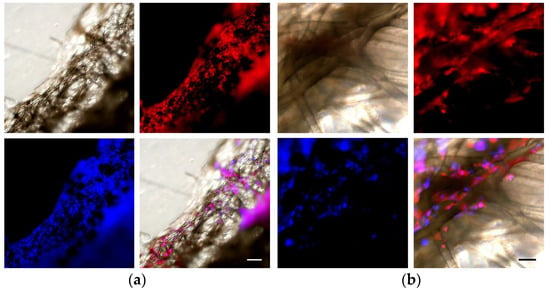
The in vitro cell culture results showed that after 1 month of culture, normal human dermal fibroblasts (NHDFs) were able to grow and proliferate well on the prepared MEW scaffolds as shown in the fluorescence microscope photographs in Figure 6. This demonstrated that the MEW scaffolds constructed using the FDM technique and cPLA molds did not induce any cytotoxicity and had good biocompatibility.
03|Application Driven Change: 3D Printing Technology Enabling Medical Innovation
The study successfully demonstrates the development of hollow microfibrous scaffolds with branching vessel bionic design by innovatively combining two additive manufacturing technologies, FDM and MEW. This strategy effectively leverages the advantages of both technologies and demonstrates great potential in the field of tissue engineering micro- and nanofabrication, while providing new solutions for personalized medicine.
Comprehensive test results show that the prepared scaffolds not only possess good biocompatibility, but also exhibit the required anisotropic mechanical strength, which meets the requirements of vascularization applications. This innovative approach not only provides a new idea for fabricating complex bionic tissue or organ structures, but also foretells a wide range of applications in future clinical medicine, especially in the application of biomedical materials and regenerative medicine.
Together with you, we look forward to the further evolution of this technology and the continued innovation and development of related technologies and medical fields. This also ties in with the core concept of the upcoming TCT2024 Asia, "Application-Driven Change", which highlights the role of real-world applications in driving 3D printing technology and medical innovation. By focusing on the technology in action, the show will not only demonstrate how an application-driven approach can facilitate the continued advancement and innovation of medical technology, but also deepen the industry's understanding and application of these transformative technologies.
Research Links:Integrating Fused Deposition Modeling and Melt Electrowriting for Engineering Branched Vasculature.DOI:10.3390/biomedicines1112313
 Home
Home Telephone
 Message