


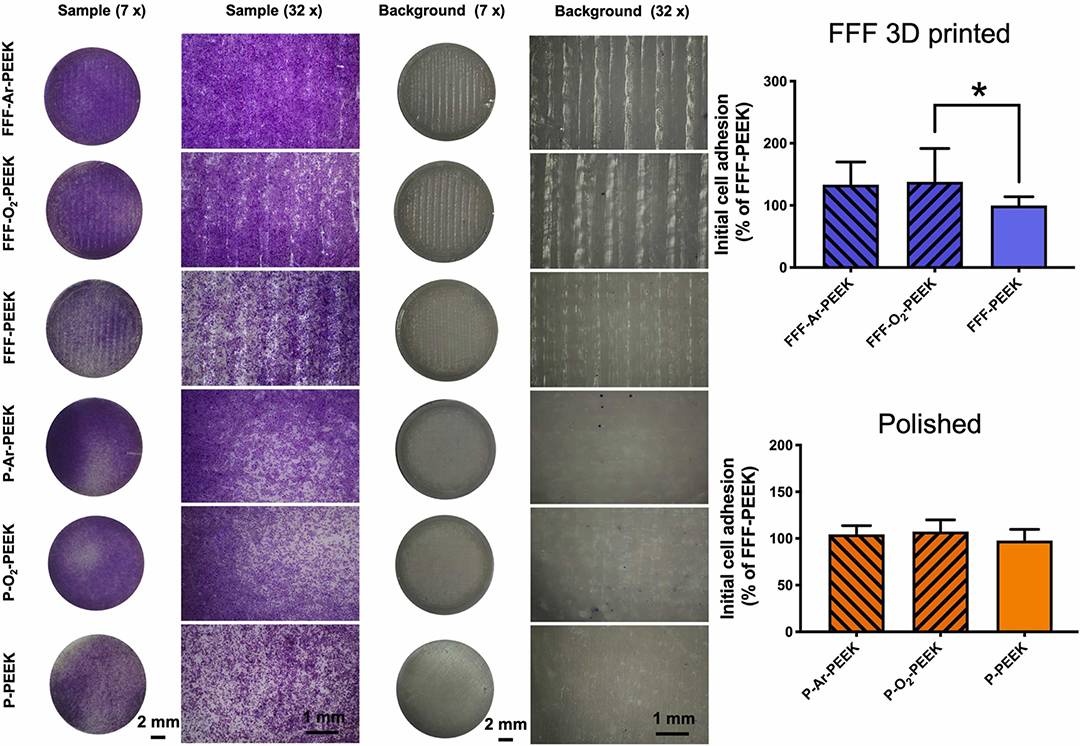

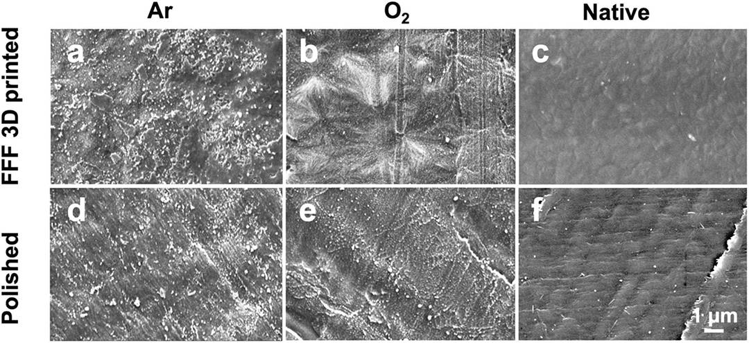
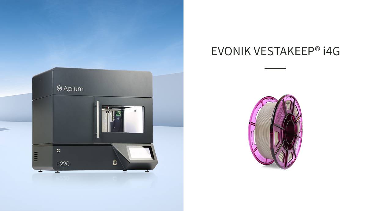


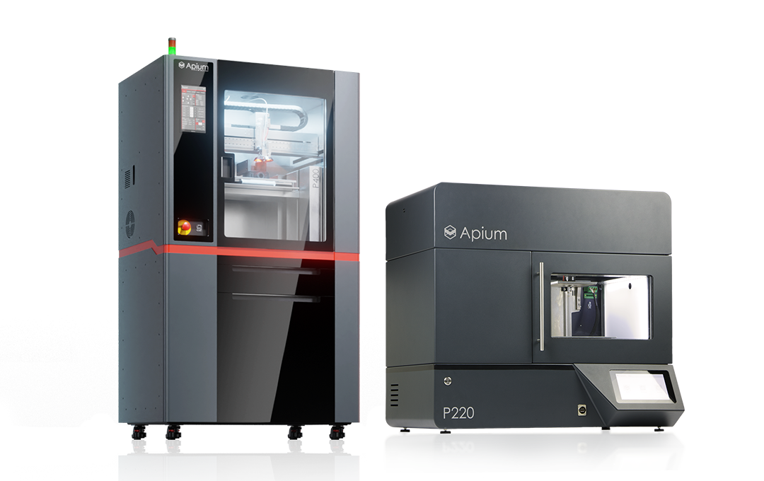
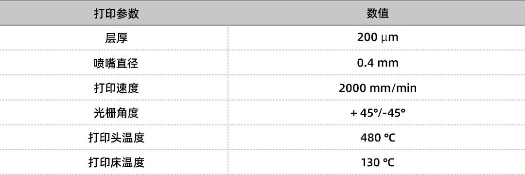
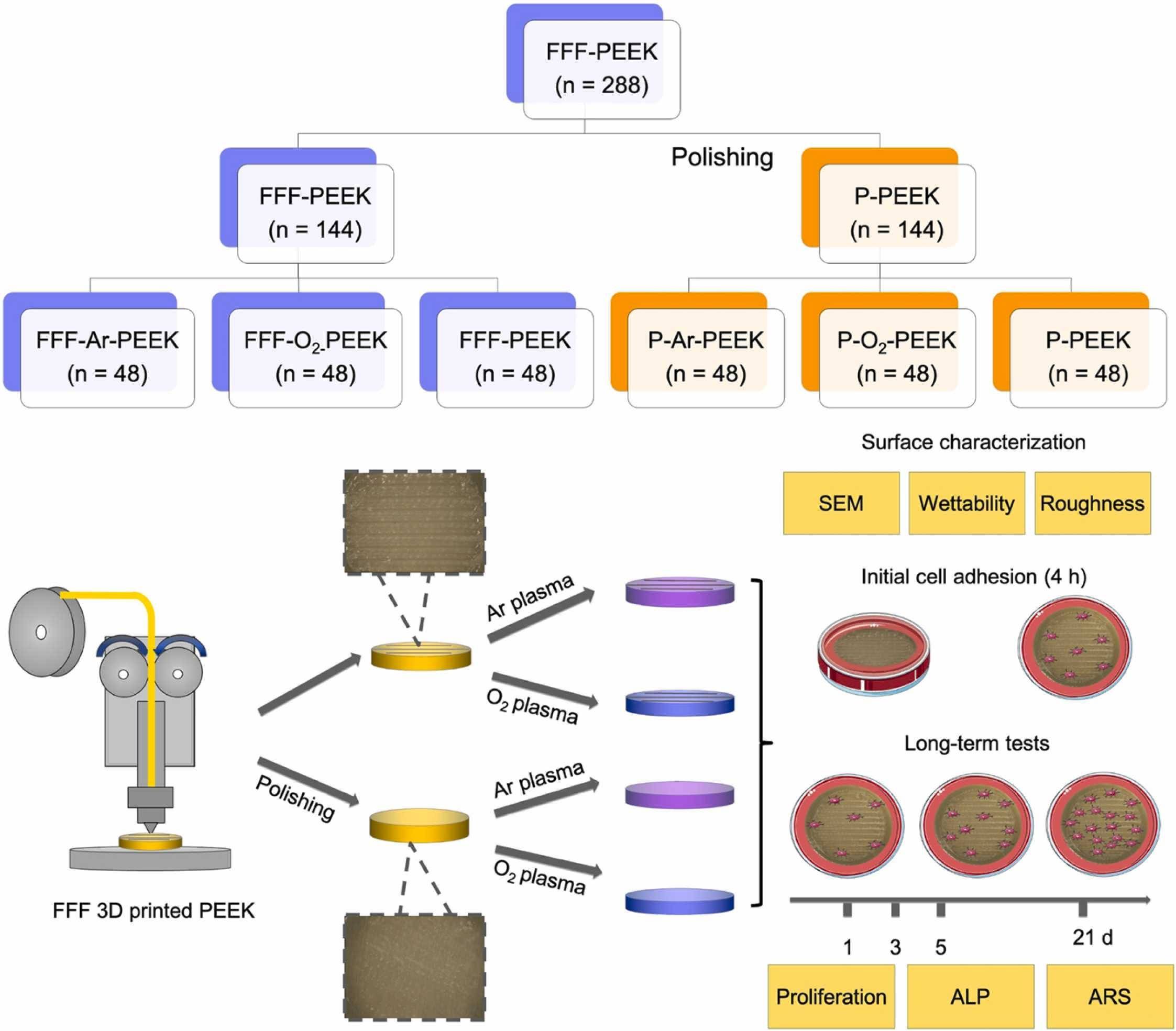
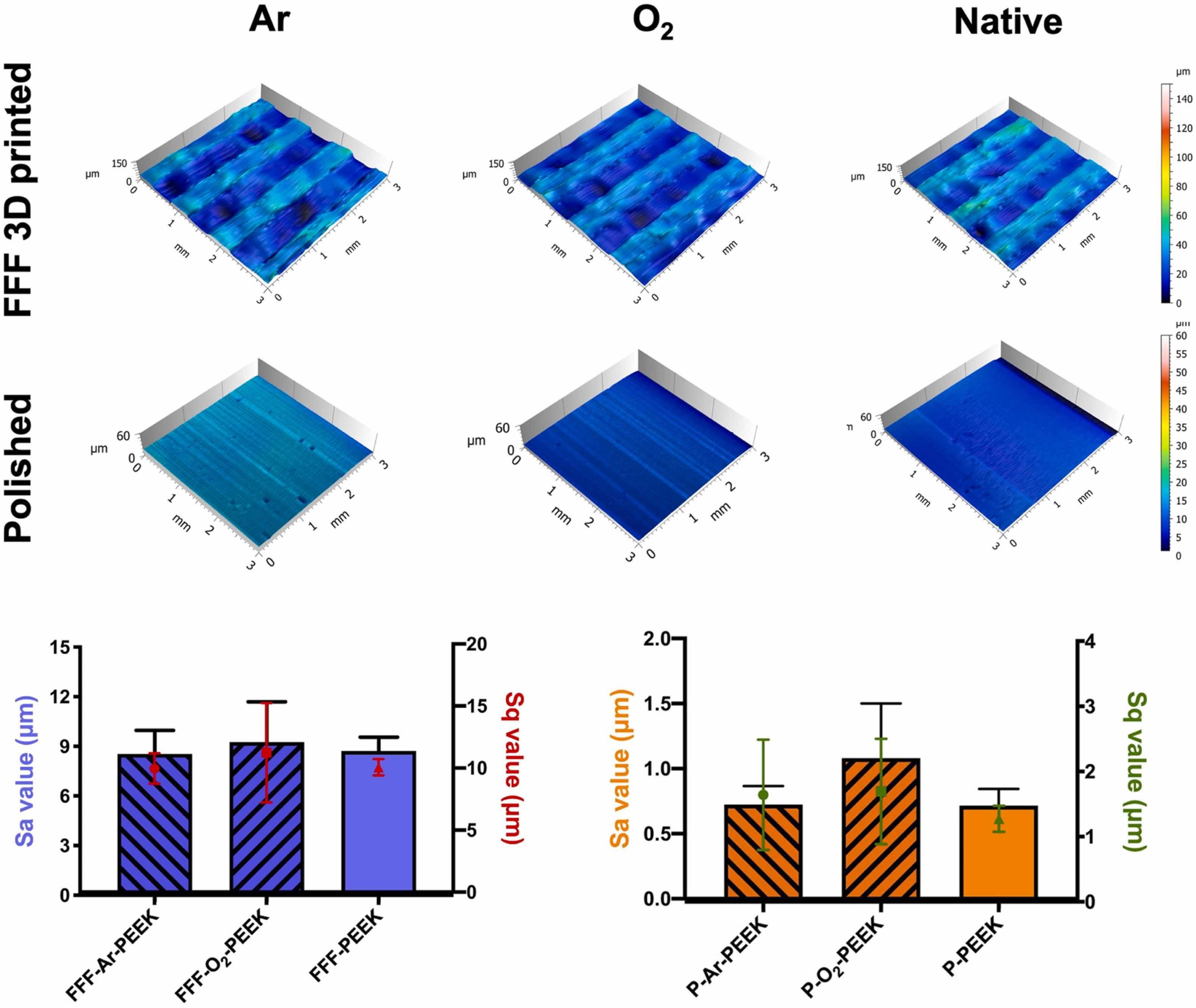
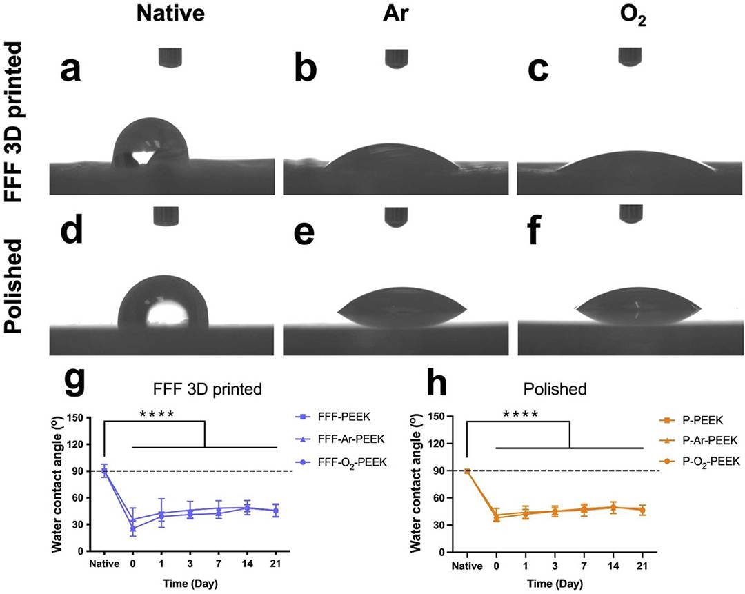
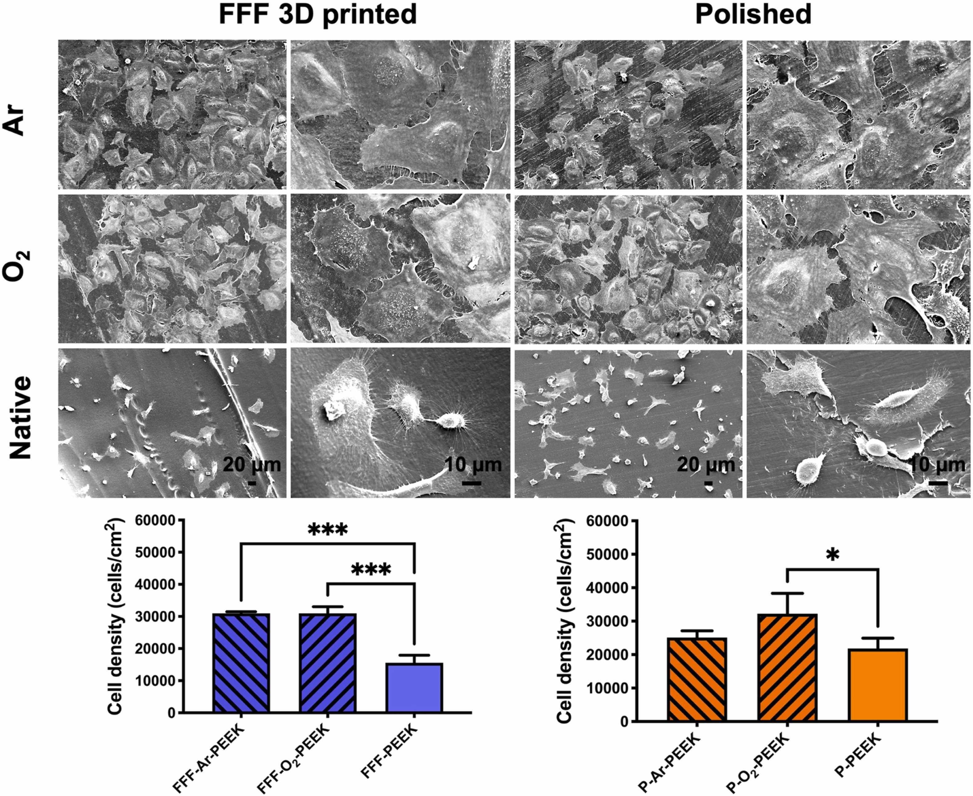
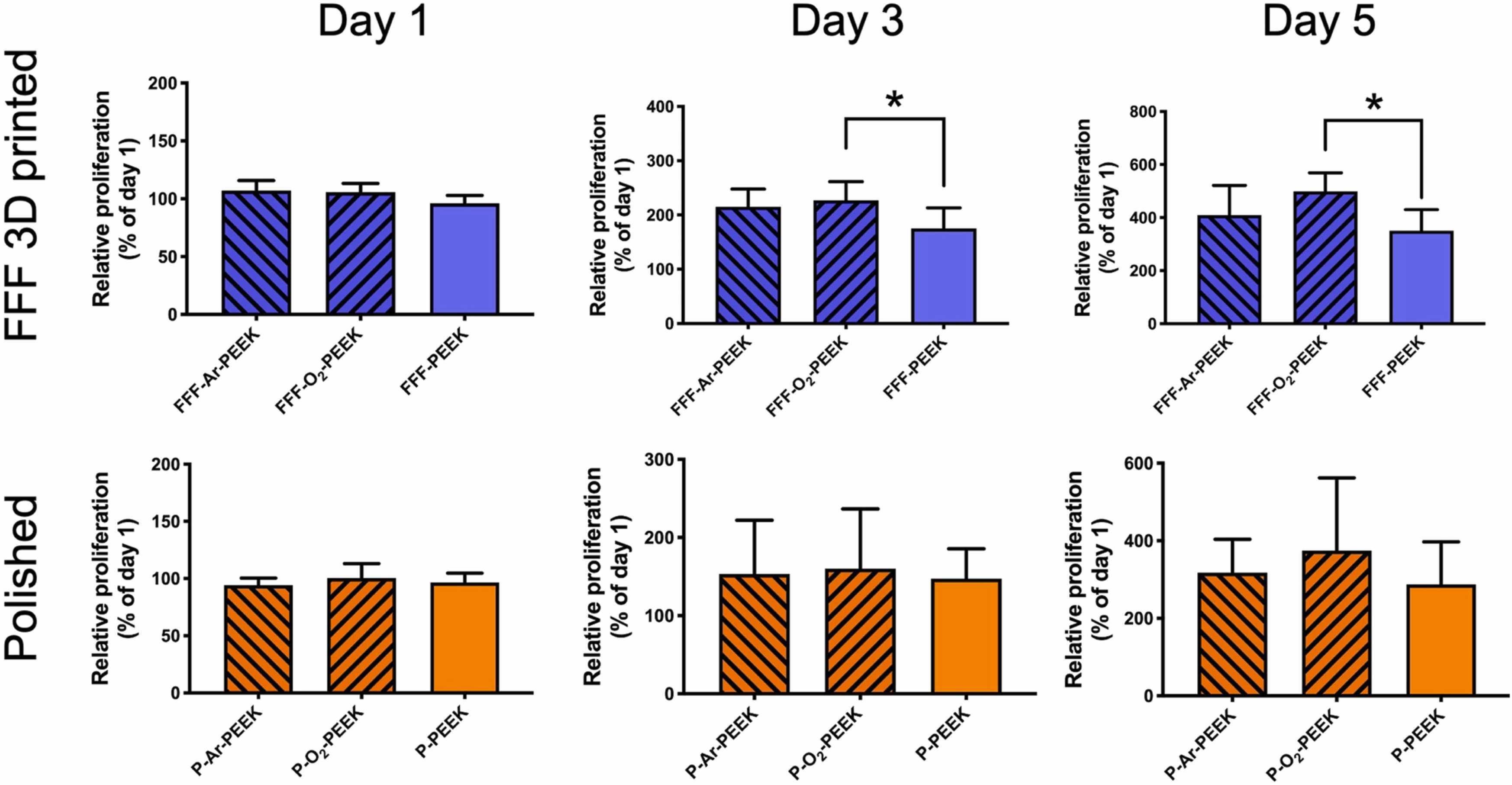
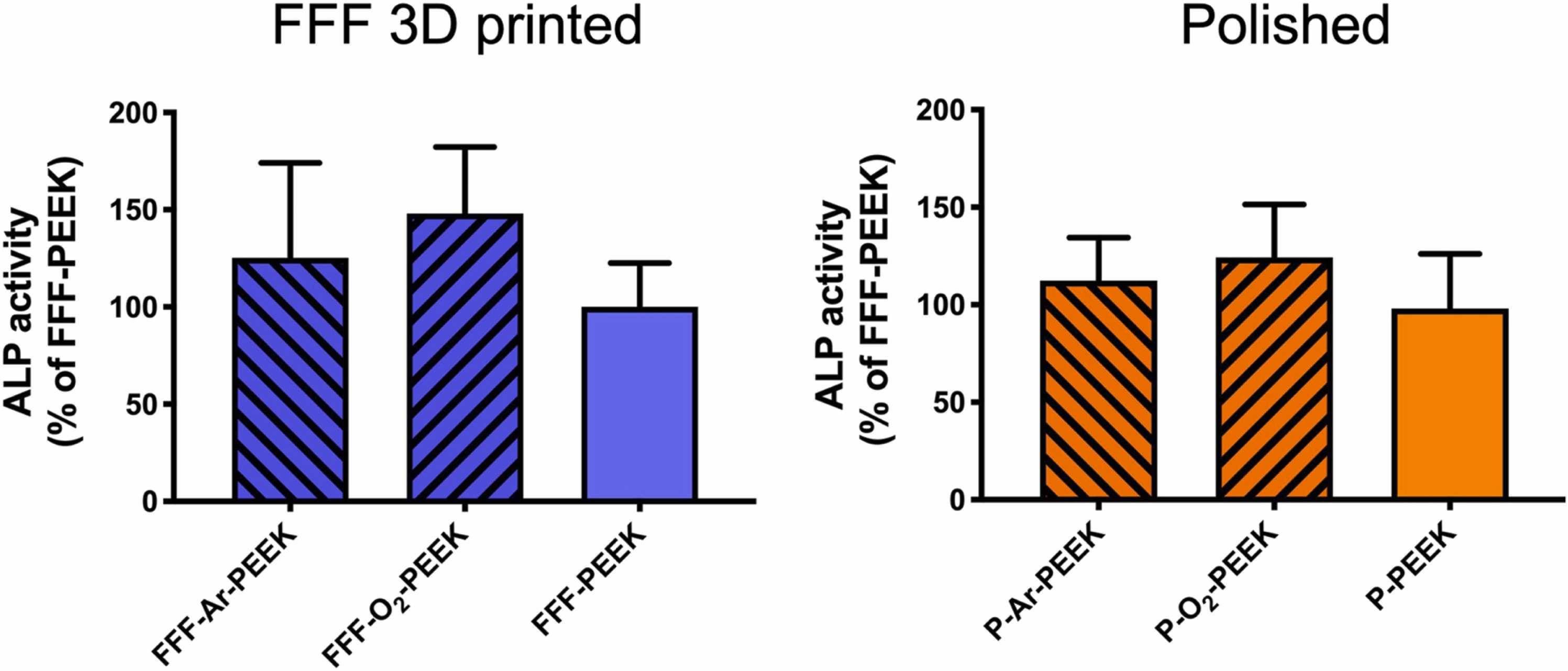
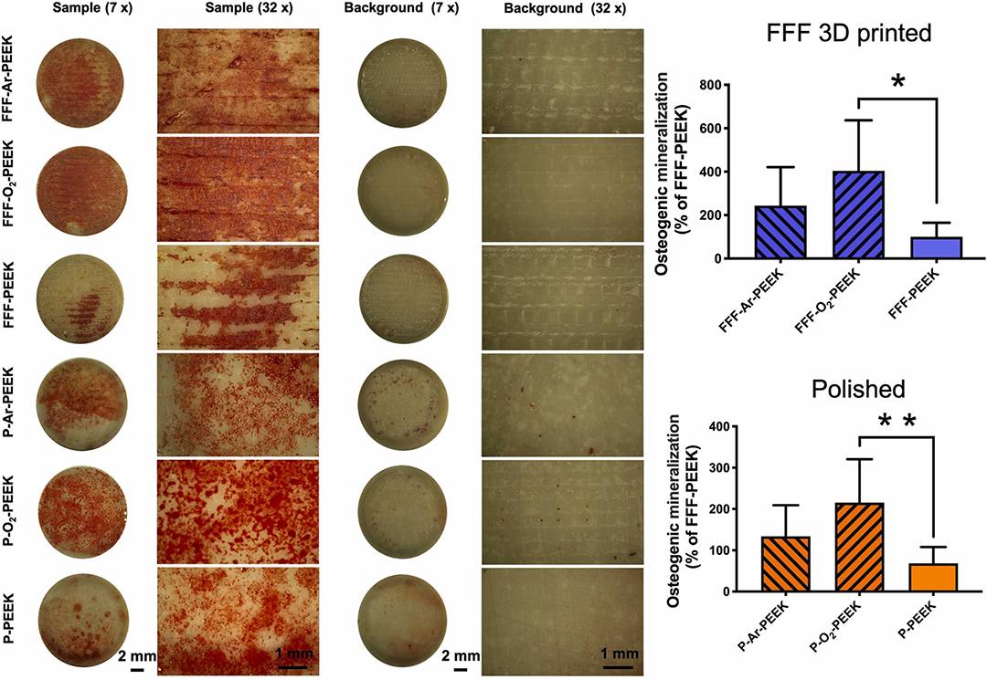
Material and Methods In this study, polyether ether ketone (PEEK) implant samples were successfully prepared using an Apium P220 FFF 3D printer. The printing parameters are shown in the table below, and medical grade PEEK 3D printing consumables (medical grade vonik VESTAKEEP®i4 G consumables) were used as the printing material. Apium P220 3D printer and Evonik VESTAKEEP®i4 G consumables. The printing parameters of the Apium P220 3D printer are shown in the table below: The printed PEEK samples were divided into two groups: polished and unpolished. The polished group samples were treated with sandpaper for polishing and the unpolished group retained the original surface properties of the 3D prints. The two groups of samples were treated by argon (Ar) and oxygen (O₂) plasma treatment (see Figure 1), with the aim of exploring the effect of plasma treatment on the surface properties of PEEK. The changes in surface morphology and wettability of the two groups of samples before and after plasma treatment were analyzed by scanning electron microscopy, contact angle measurement, and other technical means, and cell culture experiments were performed to examine the effect of plasma treatment on the in vitro osteogenic activity of the samples. Figure 1. Schematic representation of the grouping and experimental procedure. Results and Discussion 1. Changes In Surface Microstructure After argon (Ar) and oxygen (O₂) ionomer treatment, nanoscale spherical particles were formed on the surface of the PEEK samples (Figure 2). Figure 2. SEM images of FFF 3D printed and polished PEEK before and after Ar and O₂ plasma treatment at 10,000× magnification. The observed increases in the surface roughness parameters Sa and Sq were not statistically significant compared to the original samples, indicating that the plasma treatment improved the surface morphology at the microscopic level without significantly altering its macroscopic roughness (Figure. 3). Figure. 3. 3D surface topology reconstruction. 2、Improvement Of Surface Hydrophilicity The water contact angle (WCA) measurements of FFF-PEEK samples decreased from 90.4±7.4° to 35.7±13.0° after Ar treatment and 25.6±8.8° after O₂ treatment, and the WCA measurements of P-PEEK samples decreased from 89.5±2.5° to 41.1±7.3° after Ar treatment and 38.0±3.1° after O₂ treatment, and these changes were all were statistically significant (p<0.0001). As the storage time increased, although the water contact angle recovered, the measured values after 21 days were still significantly lower than the pre-treatment level, stabilizing below 60°, as shown in Figure 4. Figure 4. Water contact angle (WCA) measurements of FFF 3D printed and polished PEEK before plasma treatment and at different time points (0, 1, 3, 7, 14 and 21 days). 3. Enhancement Of Cell Adhesion After argon (Ar) and oxygen (O₂) plasma treatment, significant microstructural changes were observed on the surface of FFF-PEEK versus polished PEEK samples, especially the formation of spherical nanoparticles (shown in Figure 2). This treatment enhanced the adhesion of Saos-2 osteoblasts on the PEEK surface. after 4 h of incubation, the O₂ plasma-treated FFF-PEEK group exhibited a higher cell adhesion density compared to the untreated original samples (Figure 5). Figure. 5. Qualitative and quantitative results of initial cell adhesion after 4 hours of incubation. Continuing the incubation up to 24 h, the cells on the plasma-treated samples not only increased in number, but also had a more expanded morphology and an increased number of pseudopods (Figure. 6). Figure 6.SEM images: qualitative and quantitative observation of Saos-2 osteoblast morphology before and after plasma treatment of FFF-PEEK and P-PEEK. 4. Enhanced Cellular Metabolic Activity Plasma treatment increased the metabolic activity of Saos-2 osteoblasts on the FFF 3D printed PEEK surface (Figure. 7), and the O₂ plasma-treated FFF-PEEK samples showed an increase in cellular metabolic activity compared to untreated pristine samples during the incubation periods of days 3 and 5. Fig. 7. Comparison of relative cell metabolic activity and proliferation of Saos-2 osteoblasts before and after plasma treatment of FFF-PEEK and P-PEEK samples. 5. Changes In Alkaline Phosphatase (ALP) Activity The ALP activity of Saos-2 cells was slightly elevated after O₂ plasma treatment of the PEEK samples (Fig. 8).After 5 days of incubation, the ALP activity of the FFF-PEEK and P-PEEK groups treated with O₂ plasma increased slightly in comparison to the original samples, but this effect was not significant in the short term. Figure 8. Alkaline phosphatase (ALP) activity of Saos-2 osteoblasts after 5 days of incubation of FFF 3D printed and polished PEEK samples after plasma surface treatment versus untreated. Data are expressed as mean ± standard deviation. 6. Promotion Of Mineralized Nodule Formation The number of mineralized nodules of Saos-2 cells on PEEK samples treated with O₂ plasma was significantly increased (Fig. 9).After 21 days of incubation, the number of mineralized nodules formed by Saos-2 cells on FFF 3D printed and polished PEEK samples treated with O₂ plasma was significantly increased, suggesting an increase in the amount of calcium phosphate deposition, especially in the FFF-O₂-PEEK group. It was about 60% higher than the untreated FFF-PEEK group. Figure 9. Qualitative and quantitative results of osteogenic differentiation after 21 days of culture. Findings Taken together, the combination of plasma surface treatment technology combined with FFF 3D printing technology provides an effective way for surface modification of PEEK implants. By modulating the surface morphology and hydrophilicity of PEEK, it successfully enhanced its bioactivity and promoted the adhesion, alkaline phosphatase (ALP) activity, metabolic activity and osteogenic differentiation of Saos-2 cells. This study not only provides new technical support for the application of PEEK materials in orthopedics and dentistry, but also provides important ideas for further optimizing the biocompatibility and osseointegration ability of implants. Original Link:Tailoring the biologic responses of 3D printed PEEK medical implants by plasma functionalization.DOI:10.1016/j.dental.2022.04.026


 Home
Home Telephone
 Message